Right Shoulder Anatomy Diagram
Right Shoulder Anatomy Diagram. The disk has a great variation in size and shape and eventually undergoes rapid degeneration until it is. Posted on december 13, 2018december 12, 2018. The glenohumeral joint has the following supporting structures Last update february 25, 2021. I sustained fractures to the right shoulder & top of arm in 2003. Select from premium shoulder anatomy images of the highest quality. The human shoulder is made up of three bones:
Webmd's shoulder anatomy page provides an image of the parts of the shoulder and describes its function, shoulder problems, and more. This acts as the bony framework by which the muscles of the chest, upper back and shoulder connect the upper limb to the trunk of the body and control it's movements.the clavicle connects to the sternum via the. Mri, radiographical, and illustrated anatomical. An understanding of the anatomy of the rtc tendons and the underlying pathogenesis aids in the diagnosis, which is based. We'll remove the humerus and we'll take a look at the glenoid cavity. Blank head and neck muscles diagram | body muscles … from i.pinimg.com. Three bones come together at the shoulder joint. Besides big lifting jobs, the shoulder joint is also responsible for getting the hand in the right position for any function.
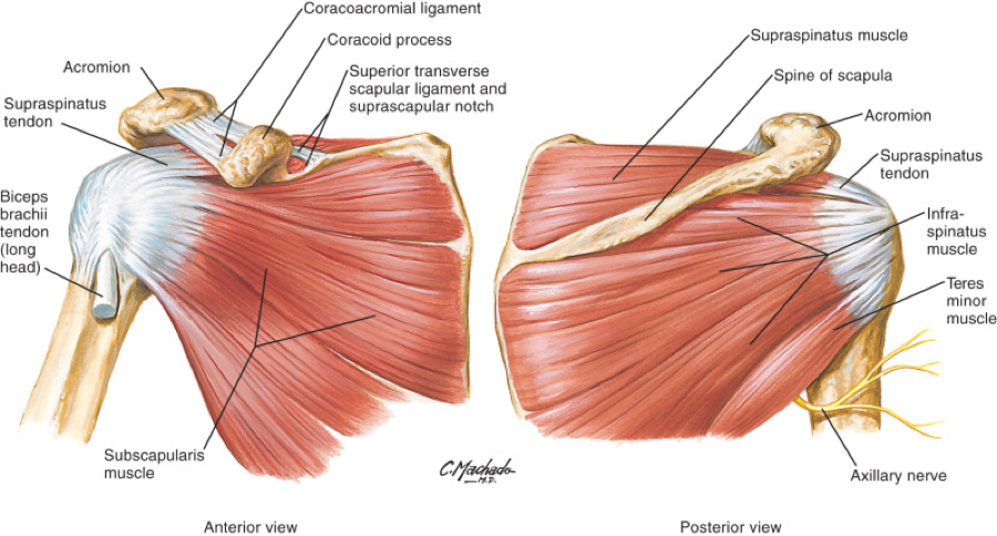
2.2 shoulder muscles and shoulder tendons.
The human shoulder is made up of three bones: Blank head and neck muscles diagram | body muscles … from i.pinimg.com. Shoulder dislocation treatment recovery symptoms. The shoulder is one of the largest and most complex joints in the body. Normal anatomy, variants and checklist. We're looking laterally now at the right shoulders. Learn more about the shoulder joint anatomy. This acts as the bony framework by which the muscles of the chest, upper back and shoulder connect the upper limb to the trunk of the body and control it's movements.the clavicle connects to the sternum via the. This diagram here just shows the joint capsule itself. Chevy impala radio wiring diagram. This flexibility allows you to hit a backhand swing in tennis or stretch to reach something on a top shelf. Shoulder radiology & anatomy at usuhs.mil. Sechrest, md narrates an animated tutorial on the basic anatomy of the shoulder. Anatomical diagram of the muscles of the neck. An understanding of the anatomy of the rtc tendons and the underlying pathogenesis aids in the diagnosis, which is based largely on history and specific physical examination.
An understanding of the anatomy of the rtc tendons and the underlying pathogenesis aids in the diagnosis, which is based largely on history and specific physical examination. The shoulder joint (glenohumeral joint) is a ball and socket joint between the scapula and the humerus. This set is often saved in the same folder as. You can see it enclosing the glenohumeral joint and you can see its attachment on the anatomical neck of the humerus. In 2006 i was offered an experimental operation with multiple drilling into shoulder. The shoulder joint is the connection between the chest and the upper extremity. In this episode of eorthopodtv, orthopaedic surgeon randale c. Our website is not intended to be a substitute for professional medical advice, diagnosis, or treatment.
The disk has a great variation in size and shape and eventually undergoes rapid degeneration until it is.
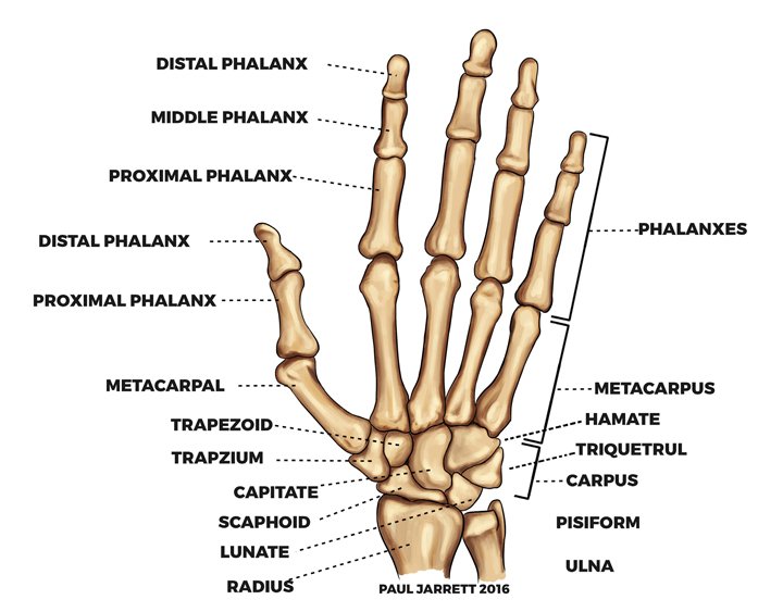
When you realize all the different ways and positions we use our hands. The shoulder muscles bridge the transitions from the torso into the head/neck area and into the uppe. The transverse humeral ligament is not shown on this diagram. This set is often saved in the same folder as. The clavicle and scapula form the shoulder girdle. Blank head and neck muscles diagram | body muscles … from i.pinimg.com. The scapula (shoulder blade), clavicle (collarbone) and humerus. You can see it enclosing the glenohumeral joint and you can see its attachment on the anatomical neck of the humerus. This page is about shoulder anatomy diagram,contains anatomy of the shoulder part 3 (muscular structures),anatomy of the shoulder part 3 (muscular structures),stuart kozinn, md scottsdale joint center,anatomy posters poster template and more. Chevy impala radio wiring diagram. Which are the shoulder muscles and where they are located?
The clavicle and scapula form the shoulder girdle. Sechrest, md narrates an animated tutorial on the basic anatomy of the shoulder. 2.1 bones of the shoulder girdle. Anatomical diagram of the muscles of the neck. Image result for glenohumeral ligaments right shoulder. Shoulder dislocation treatment recovery symptoms. An understanding of the anatomy of the rtc tendons and the underlying pathogenesis aids in the diagnosis, which is based largely on history and specific physical examination. Besides big lifting jobs, the shoulder joint is also responsible for getting the hand in the right position for any function. Posted on december 13, 2018december 12, 2018.

The shoulder joint is formed where the humerus (upper arm bone) fits into the scapula.
This set is often saved in the same folder as. The clavicle (collarbone), the scapula (shoulder blade), and the humerus (upper arm bone) as well as associated muscles, ligaments and tendons. Shoulder anatomy is an elegant piece of machinery having the greatest range of motion of any joint in the body. Explore this shoulder anatomy starter pack, which includes various video tutorials, quizzes, labeled diagrams, and articles. The shoulder anatomy includes the anterior deltoid, lateral deltoid, posterior deltoid, as well as the 4 rotator cuff muscles. We're looking laterally now at the right shoulders. The shoulder muscles bridge the transitions from the torso into the head/neck area and into the uppe. Human anatomical atlas of the shoulder : 2.2 shoulder muscles and shoulder tendons. Three bones come together at the shoulder joint. Learn more about the shoulder joint anatomy. Posted on december 13, 2018december 12, 2018. Discover how your shoulder works. In this episode of eorthopodtv, orthopaedic surgeon randale c. An understanding of the anatomy of the rtc tendons and the underlying pathogenesis aids in the diagnosis, which is based.
Webmd's shoulder anatomy page provides an image of the parts of the shoulder and describes its function, shoulder problems, and more shoulder anatomy diagram. The scapula (shoulder blade), clavicle (collarbone) and humerus.

Posting Komentar untuk "Right Shoulder Anatomy Diagram"